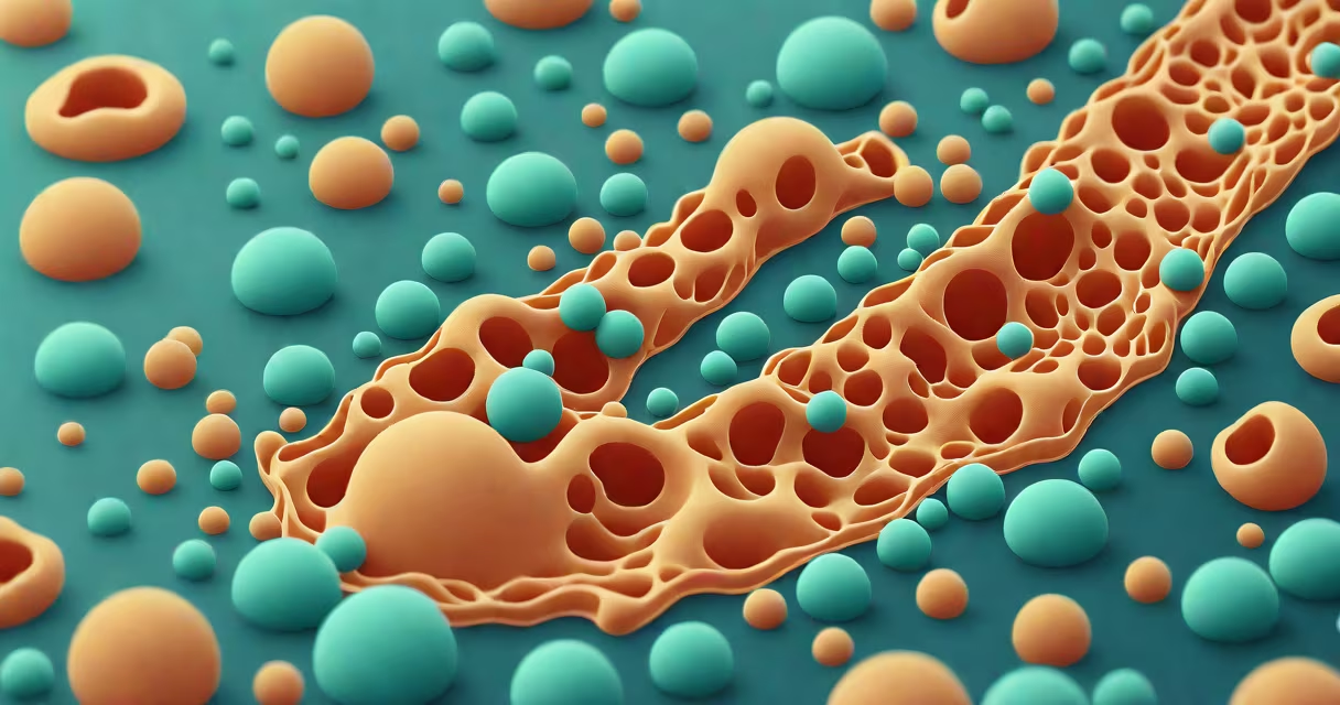
The rough endoplasmic reticulum (rER) is a network of flattened sacs in eukaryotic cells that synthesizes and modifies proteins destined for secretion, integration into membranes, or delivery to lysosomes. Its surface is studded with ribosomes, giving it a rough appearance under electron microscopy.
Proteins made by the rER begin their life at ribosomes attached to the membrane. As the polypeptide chain emerges from the ribosome, it enters the lumen of the rER through a channel called the translocon. Inside this space, the protein folds into its correct three-dimensional structure with the help of chaperone proteins like BiP and calnexin. Disulfide bonds form to stabilize the folded state, and N-linked glycosylation adds sugar groups to certain amino acids.
Once properly folded and modified, proteins are packaged into transport vesicles that bud off from the rER membrane. These vesicles travel to the Golgi apparatus, where further modifications occur. From there, they move to their final destination-either released from the cell via exocytosis, embedded in the plasma membrane, or delivered to lysosomes.
The rER consists of interconnected cisternae-flattened membrane-bound sacs-derived from the nuclear envelope. The outer membrane of the nuclear envelope is continuous with the rER membrane, allowing direct communication between the nucleus and the protein production system.
Key structural elements include:
When misfolded proteins accumulate, the rER triggers the unfolded protein response (UPR), a signaling pathway that either restores homeostasis or initiates apoptosis if stress persists. This mechanism protects the cell from toxic protein aggregates.
The rER is essential for producing secretory proteins, membrane proteins, and soluble proteins destined for lysosomes. For example, insulin-a hormone critical for glucose regulation-is synthesized in pancreatic beta cells as a precursor called preproinsulin. The signal peptide directs it to the rER, where it is cleaved and folded. After modification, it moves to the Golgi and is stored in secretory granules until blood glucose levels rise.
Another example is immunoglobulin G (IgG), the most common antibody in human blood. B cells produce IgG in large quantities using rER machinery. Each heavy chain is synthesized with a signal sequence that targets it to the rER. Once inside, disulfide bridges form between chains, and glycosylation occurs. Without functional rER, these antibodies would not fold correctly and would be degraded.
| Protein Type | Destination | Example | Processing Step |
|---|---|---|---|
| Secretory | Extracellular space | Insulin | N-linked glycosylation, signal peptide cleavage |
| Membrane-bound | Plasma membrane | EGFR (epidermal growth factor receptor) | Signal anchor sequence, glycosylation |
| Lysosomal | Lysosomes | Hexosaminidase A | Manose-6-phosphate tagging |
The rough ER differs from the smooth endoplasmic reticulum (sER), which lacks ribosomes and performs distinct functions. While the rER focuses on protein synthesis and processing, the sER handles lipid synthesis, detoxification, and calcium storage.
For instance, in liver cells, the sER metabolizes drugs and alcohol through cytochrome P450 enzymes. In muscle cells, the sER (called sarcoplasmic reticulum) stores and releases calcium ions to trigger contraction. These roles do not overlap with rER activity.
A common misconception is that both ER types perform similar tasks. They don't. The presence or absence of ribosomes defines their primary function. Confusing them leads to errors in understanding cellular trafficking pathways.
Defects in rER function cause several diseases. Cystic fibrosis results from mutations in the CFTR gene, leading to misfolded CFTR protein. This defective protein accumulates in the rER and is degraded before reaching the cell surface. The resulting lack of chloride channels disrupts mucus clearance in lungs and pancreas.
Another example is Wolfram syndrome, a rare disorder linked to mutations in the WFS1 gene. The WFS1 protein normally regulates calcium levels in the rER. When dysfunctional, chronic ER stress activates apoptosis in insulin-producing beta cells, contributing to diabetes onset.
One non-obvious fact: the rER is not static. It dynamically remodels during the cell cycle. During mitosis, it fragments and reassembles after cytokinesis. This remodeling ensures proper distribution of protein synthesis capacity to daughter cells.
I observed this fragmentation firsthand while studying dividing HeLa cells under cryo-electron tomography. The rER broke into small vesicles during metaphase, then reformed into sheets and tubules after anaphase. This process is regulated by the protein kinase GSK3β and the ER-shaping protein REEP5.
Many assume the rER produces all proteins in the cell. That's incorrect. Cytoplasmic ribosomes synthesize proteins for use within the cytosol, mitochondria, or nucleus. Only proteins with a signal peptide are targeted to the rER.
Also, the rER does not modify proteins after export. Final modifications happen in the Golgi. The rER only prepares proteins for transport. Mistaking this step leads to confusion about intracellular trafficking routes.
The rough endoplasmic reticulum is central to protein biogenesis. It ensures quality control, proper folding, and directed trafficking. Without it, multicellular life as we know it would not exist.